RESOURCE PAGE
This collection of resources are designed to give an overview of the AnatomySCAPES educational focus. The following are included for your learning:
- Fascia Research – a collection of links to foundational and relevant research articles on fascia that we reference in our classes. Most of them are open access.
- AnatomyBRIEFS – a series of short films shot on location in the AnatomySCAPES research and educational labs in San Diego, CA. The dissections highlight our focus on fascial anatomy and tissue relationships.
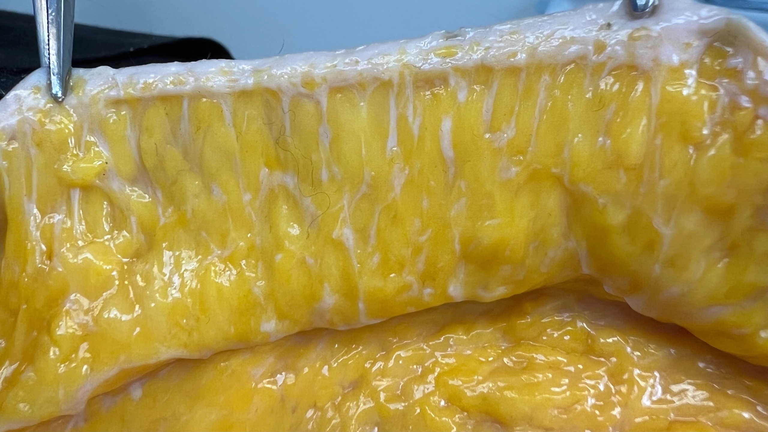
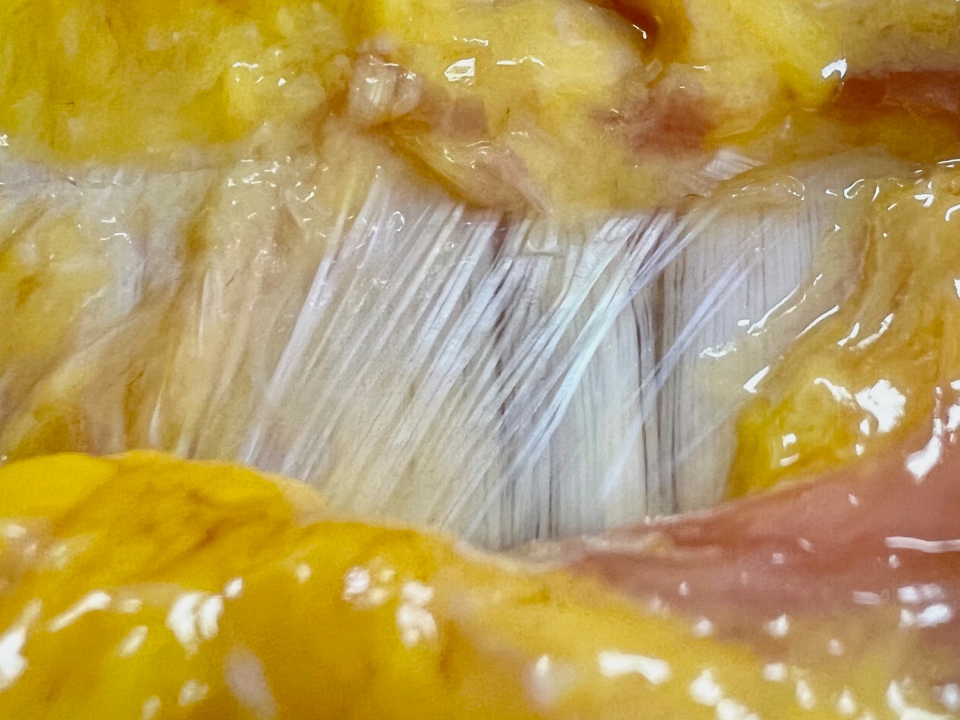
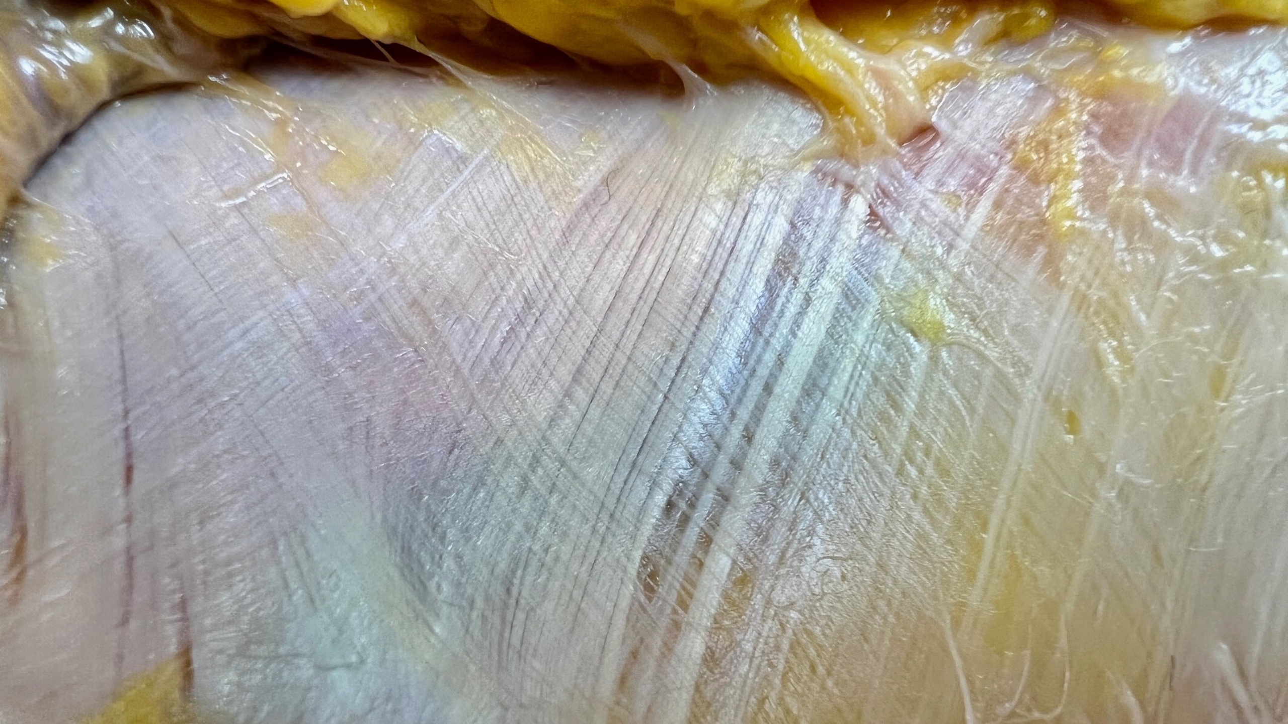
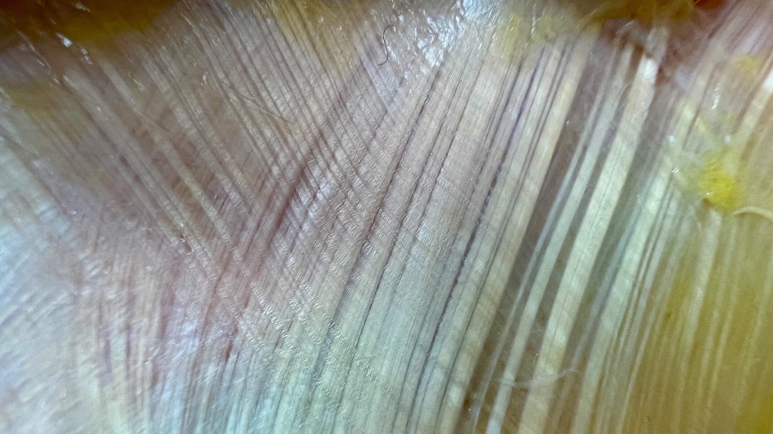
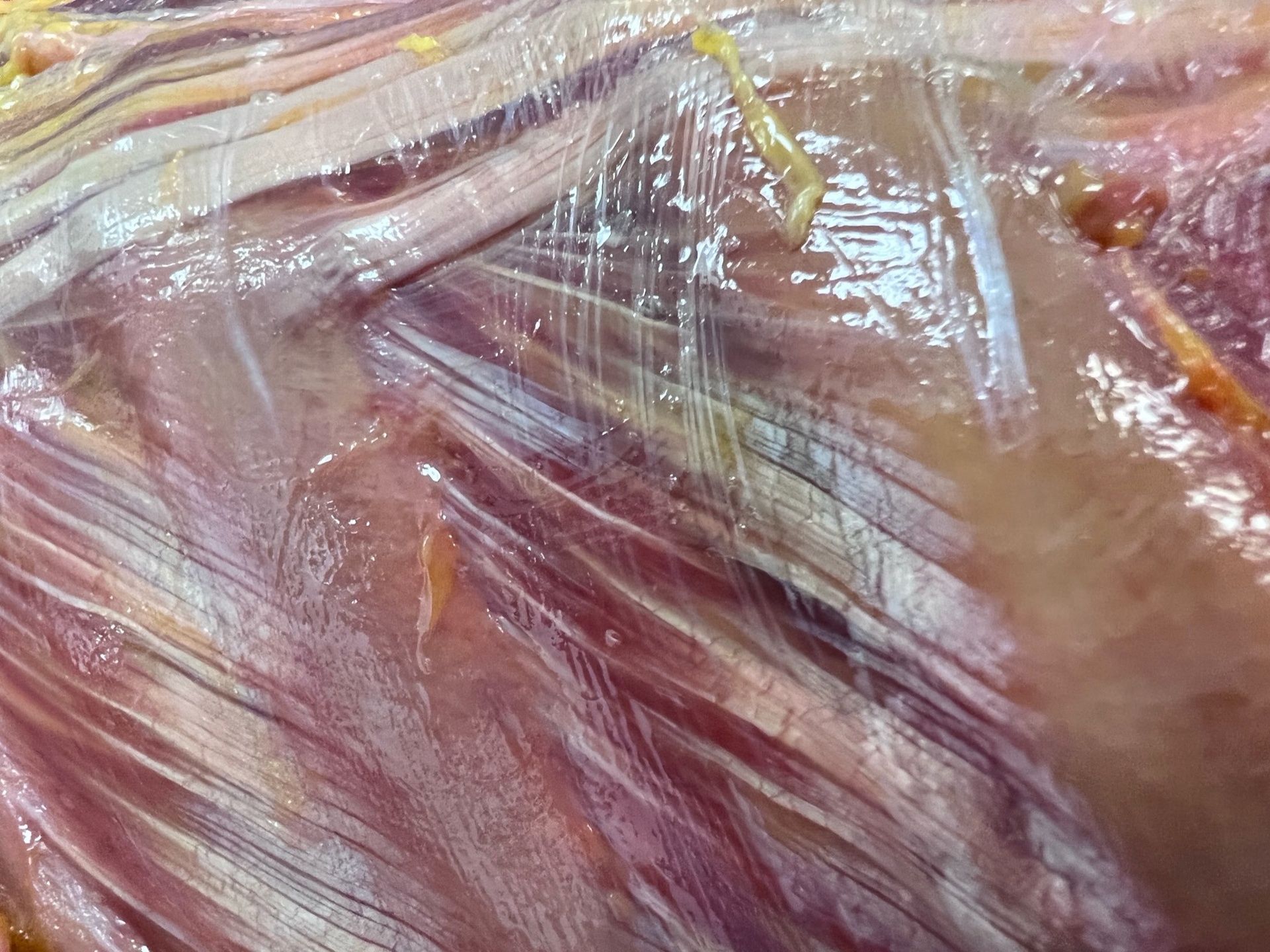
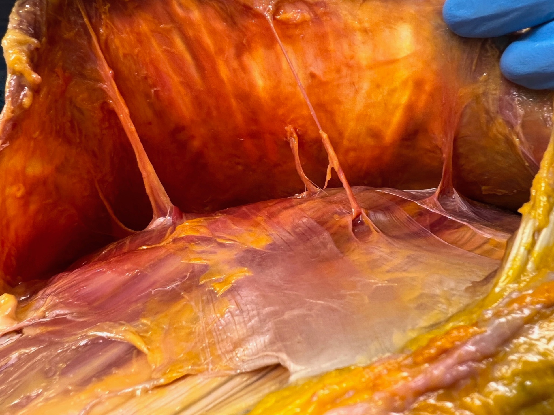
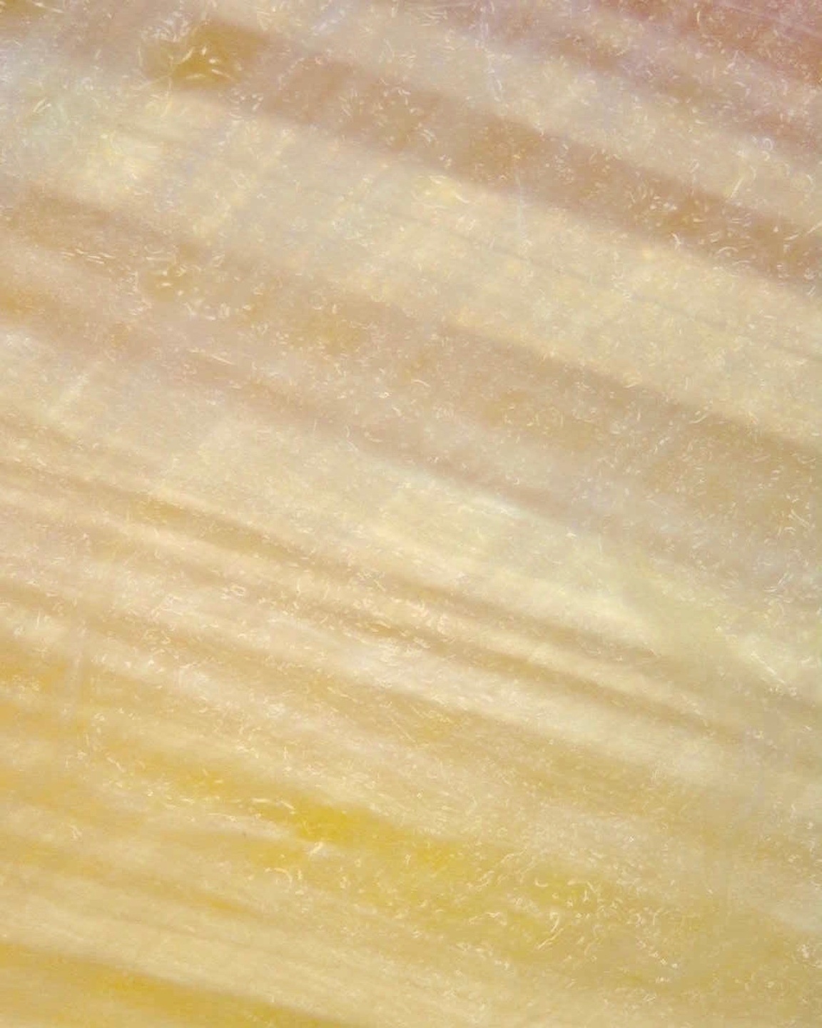
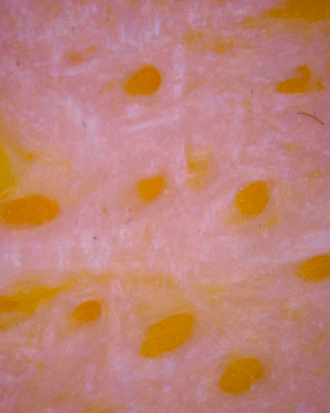
- Dissection Images – a collection of images from the dissection lab that highlight fascial tissue architecture and relationships.
CONNECT WITH US!
[email protected]
Nicole Trombley & Rachelle Clauson
Co-Directors, AnatomySCAPES, LLC

In Gratitude. . .
The images from the anatomy lab would not have been possible without the gracious gifts of the donors and their families to whom we are deeply grateful.
©2021-2026 AnatomySCAPES
All content is protected under copyright law and is the sole property of AnatomySCAPES, LLC.All rights reserved. No portion of this presentation may be published, duplicated, or edited from its original form without prior written permission from AnatomySCAPES, LLC.
Our print and digital content is a labor of love for the purpose of providing educational information to movement and bodywork professionals. If you'd like to use any of our material for your trainings, please contact us directly.
FASCIA RESEARCH
Scientific research on fascia is growing rapidly! In the early 2000s, fewer than 500 articles were published each year; today, over 1,500 per year (and growing). Below are links to foundational pieces and articles we reference in our classes. Most of them are open access.
What IS fascia?
What IS fascia? There is a lot of historical and ongoing confusion on what fascia actually is — both the Fascia Research Society's nomenclature committee and leading researchers have proposed definitions to help define fascia and facilitate communication. Here are a few papers with important proposals for helping us come to consensus on what we mean when we say "fascia." This is ongoing work! Consensus has not been reached.


Thoracolumbar Fascia
The thoracolumbar fascia is easily the most studied fascial "structure" in the human body, ranging from its biomechanical role to its likely role in nonspecific low back pain. Here, Willard, et al.'s classic review from 2012 maps out the three-dimensional anatomy of the TLF. The papers by Langevin 2011), Mense (2019), and Wilke (2017) all map out the innervation and tissue alterations that establish evidence for TLF's role in LBP. An entire generation of grad students is writing their dissertations modeled after Langevin's work!
Fascia Lata and Fascia of the Limbs
Mike Benjamin's 2009 review "The fascia of the limbs and back" is one of the most cited articles detailing the literature on the anatomy of fascia — from skin to deep, and from head to toe. The other articles are a sampling of work from Carla Stecco's research team at the University of Padua (Italy) as they continue with their aim to document the fascial system through combined means of 1) dissection, 2) histology, and 3) ultrasound.


Loose Connective Tissue, Hyaluronan & Densification


DISSECTION AnatomyBRIEFS
*NO SCREENSHOTS, SCREEN RECORDING, OR COPYING.*
AnatomySCAPES' future access to donor programs relies on our collective responsibility with these materials. The images shown here must remain contained within the educational location you are viewing it with no exceptions.
DISSECTION LAB IMAGES
*NO SCREENSHOTS, SCREENRECORDING, OR COPYING.*
AnatomySCAPES' future access to donor programs relies on our collective responsibility with these materials. The images shown here must remain contained within the educational location you are viewing it with no exceptions.