September 2026: DLI
Brittany and Chelsea!
You did a thing.
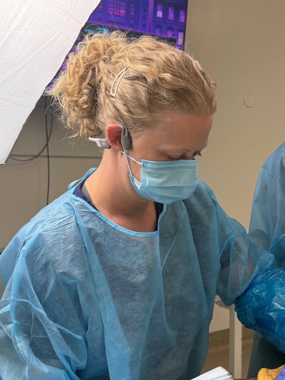
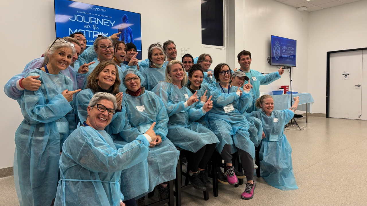
On this page, we have gathered some images and video from our week together in the lab, as well as some additional resources you might find of interest.
CANDIDS: You are welcome to grab, download, share. Please tag us on social media! @anatomyscapes
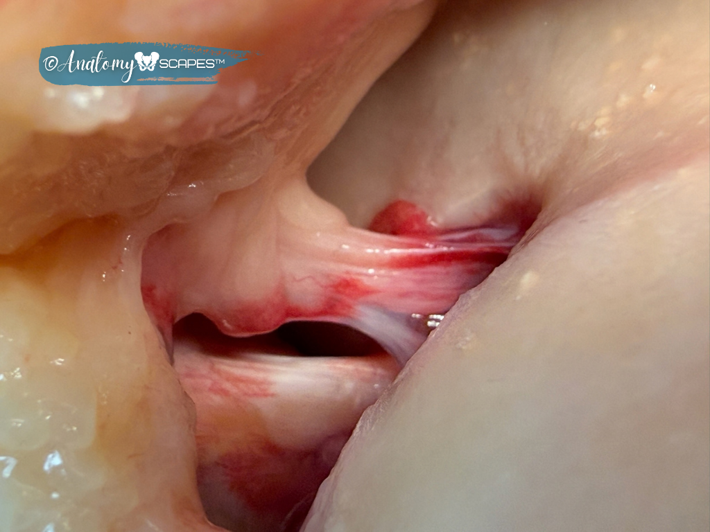
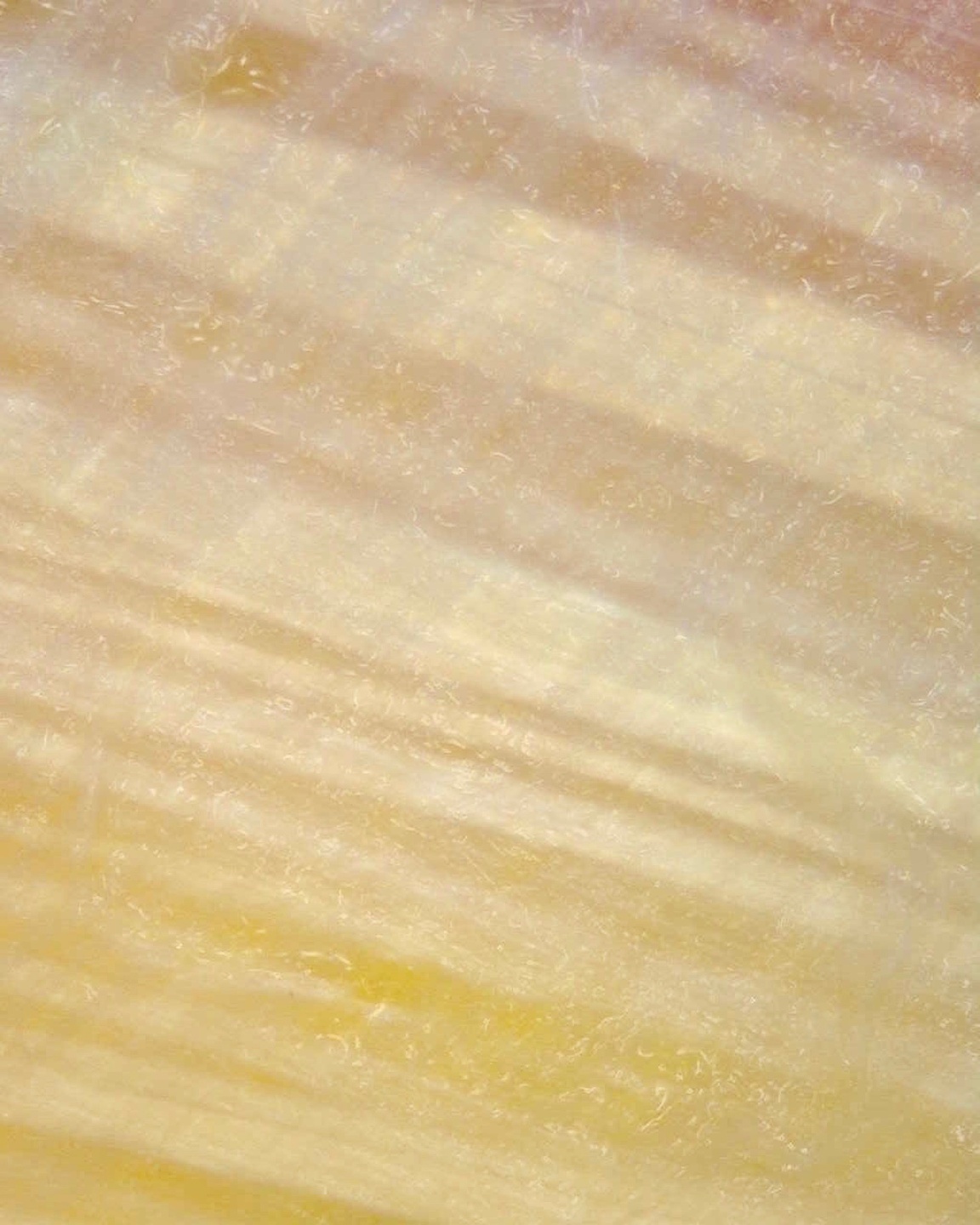
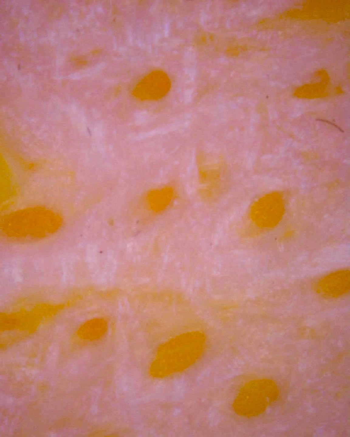
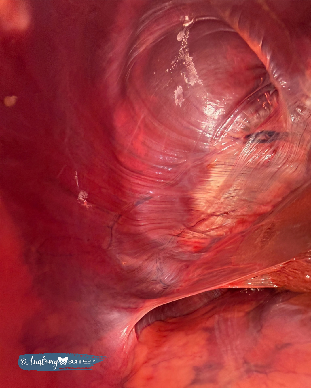
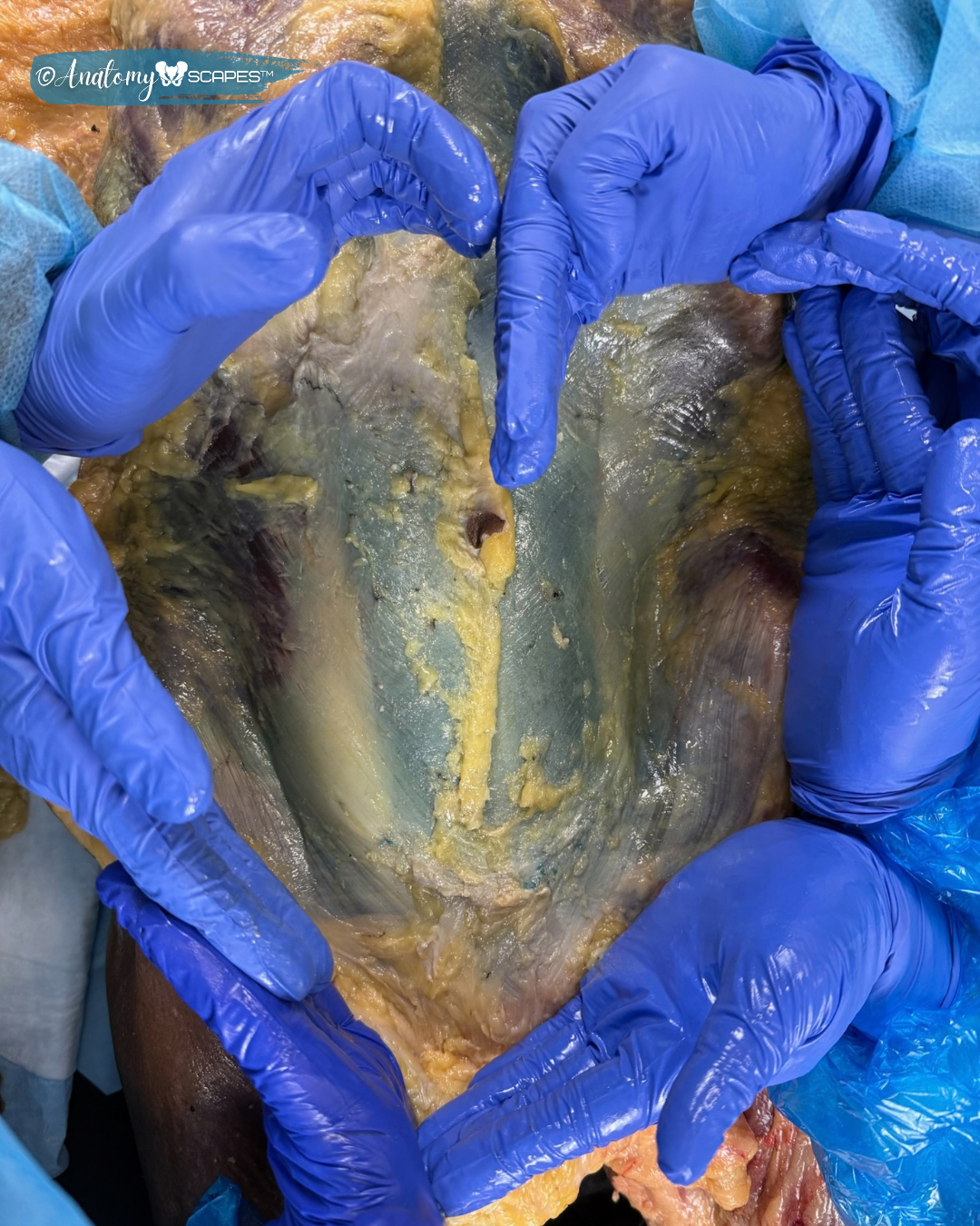
DISSECTION IMAGERY & VIDEO: These images must stay on this page! No copying or sharing! Thanks for helping us protect donor privacy AND our ongoing access to the dissection lab.
Stay in touch and we hope to see you back in the lab sometime soon!
Love,
Rachelle & Nicole

In Gratitude. . .
The images from the anatomy lab would not have been possible without the gracious gifts of the donors and their families to whom we are deeply grateful.
Welcome to the DISSECTION LAB INTENSIVE





FASCIA RESEARCH
Scientific research on fascia is growing rapidly! In the early 2000s, fewer than 500 articles were published each year; today, over 1,500 per year (and growing). Below are links to foundational pieces and articles we reference in our classes. Most of them are open access.
What IS fascia?
What IS fascia? There is a lot of historical and ongoing confusion on what fascia actually is — both the Fascia Research Society's nomenclature committee and leading researchers have proposed definitions to help define fascia and facilitate communication. Here are a few papers with important proposals for helping us come to consensus on what we mean when we say "fascia." This is ongoing work! Consensus has not been reached.


Thoracolumbar Fascia
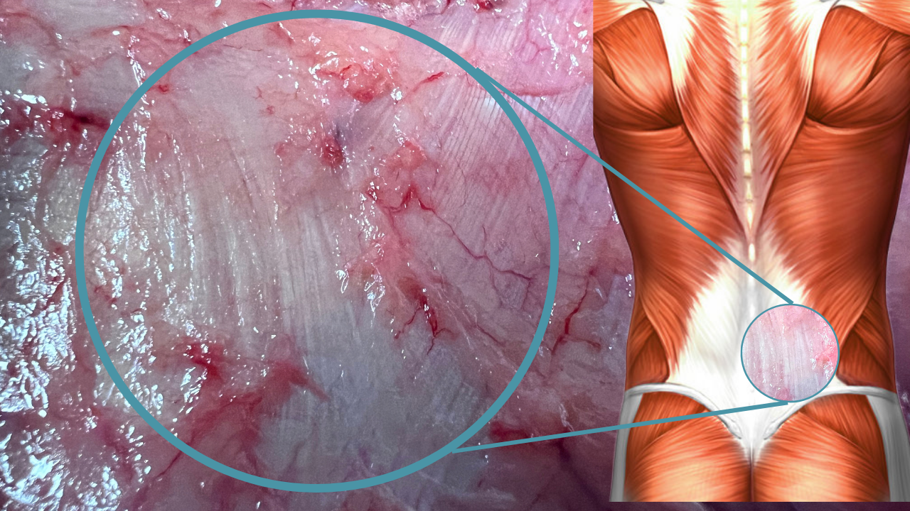
The thoracolumbar fascia is easily the most studied fascial "structure" in the human body, ranging from its biomechanical role to its likely role in nonspecific low back pain. Here, Willard, et al.'s classic review from 2012 maps out the three-dimensional anatomy of the TLF. The papers by Langevin 2011), Mense (2019), and Wilke (2017) all map out the innervation and tissue alterations that establish evidence for TLF's role in LBP. An entire generation of grad students is writing their dissertations modeled after Langevin's work!


Fascia Lata and Fascia of the Limbs
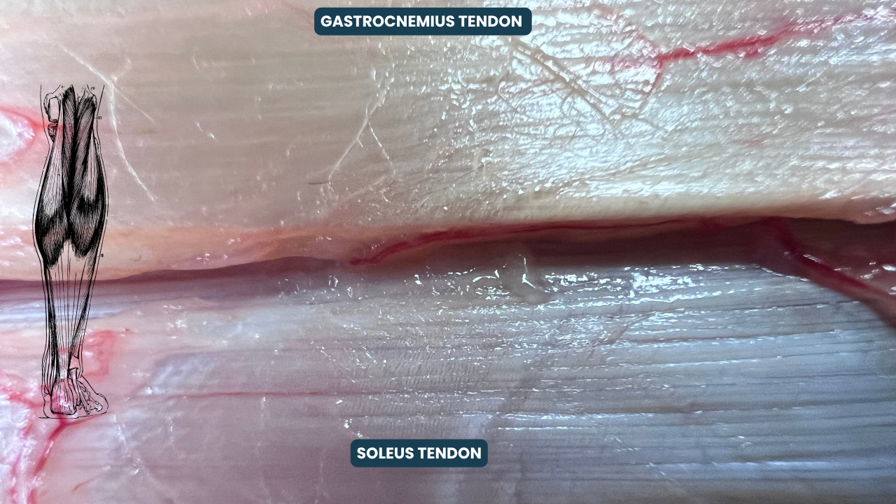
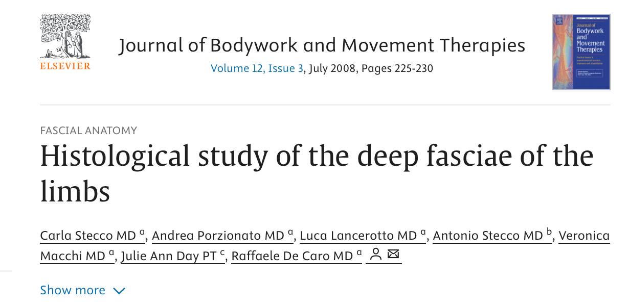
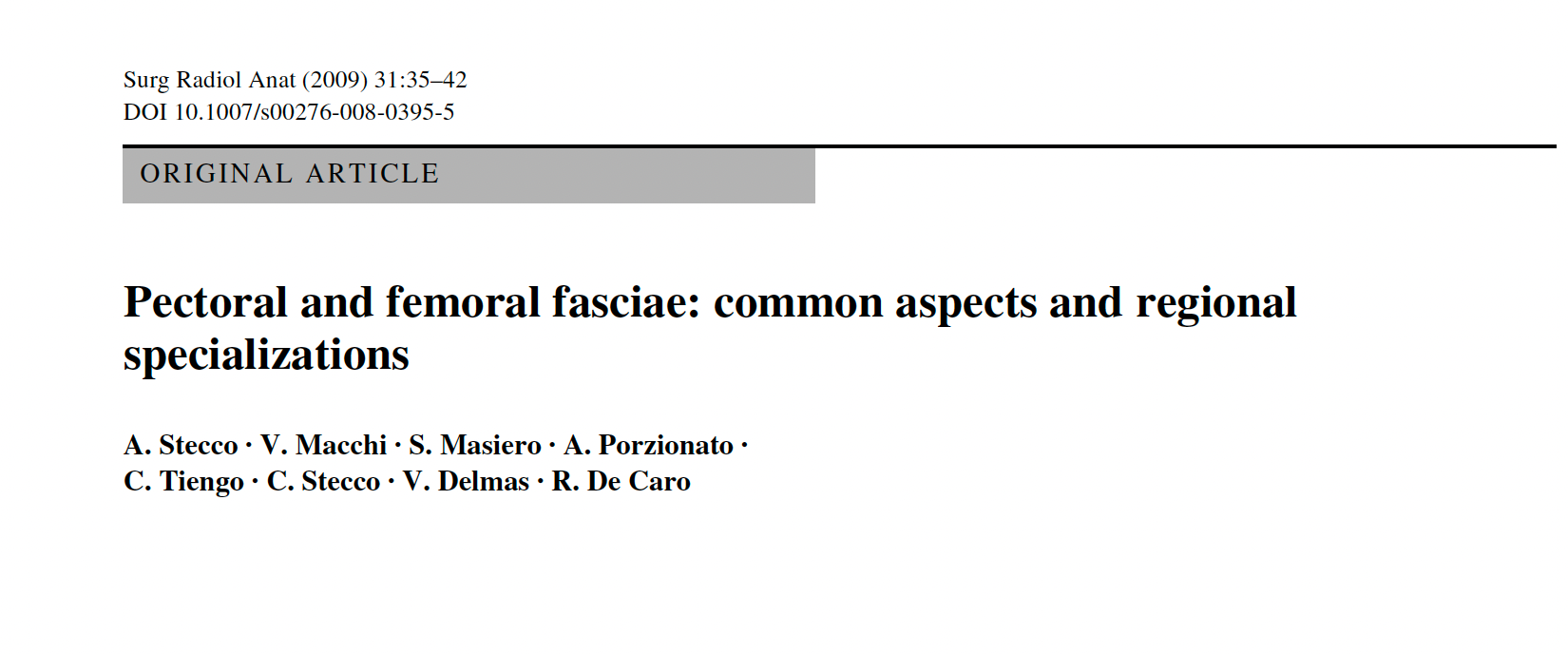
Mike Benjamin's 2009 review "The fascia of the limbs and back" is one of the most cited articles detailing the literature on the anatomy of fascia — from skin to deep, and from head to toe. The other articles are a sampling of work from Carla Stecco's research team at the University of Padua (Italy) as they continue with their aim to document the fascial system through combined means of 1) dissection, 2) histology, and 3) ultrasound.



Loose Connective Tissue, Hyaluronan & Densification


🛝GLIDE:
Skin Mobility, Subcutaneous Bursa and Fascial Specializations
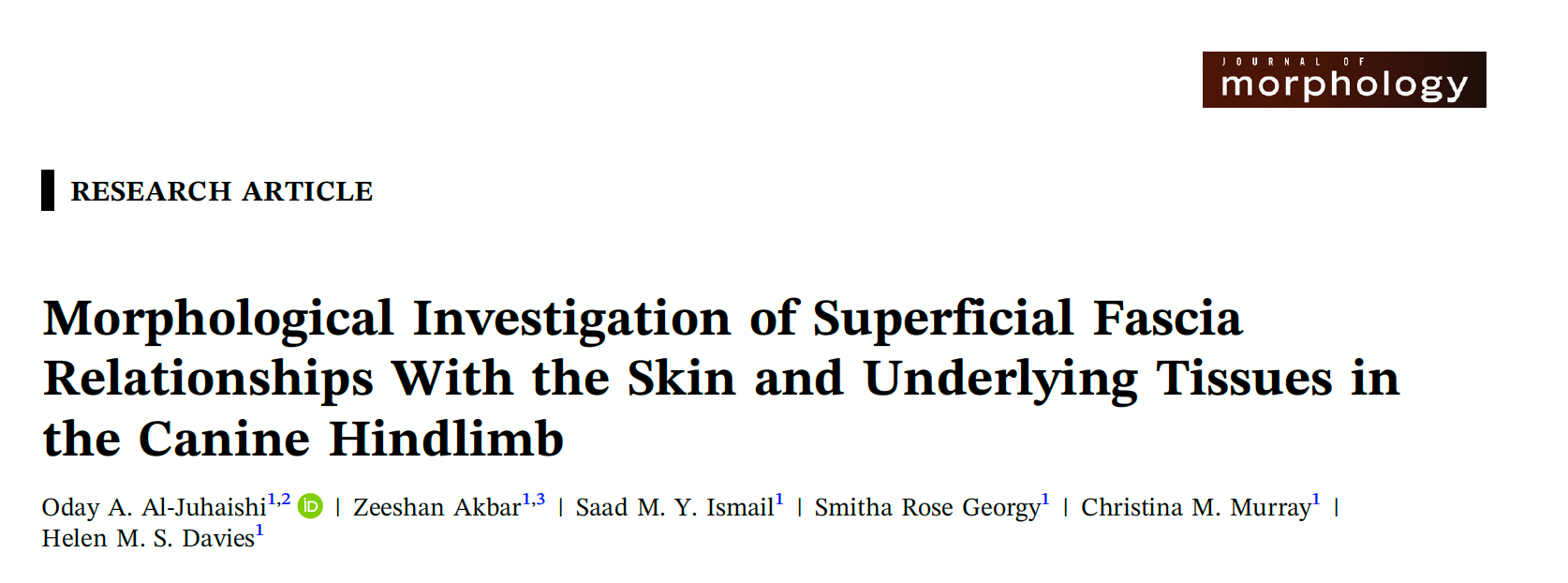
Given this year's DLI group interests in both🐕 canine fascia and 💪 olecranon process subcutaneous bursa, we've included a few more pieces to nerd out on. Both give a great understanding of these specialized SUPERFICIAL FASCIA functions.
COMPARATIVE ANATOMY: What helps your dog have such mobile, grabbable, smooshy skin? Does your dog have an olecranon bursa? What is the tissue architecture that allows for this skin mobility?
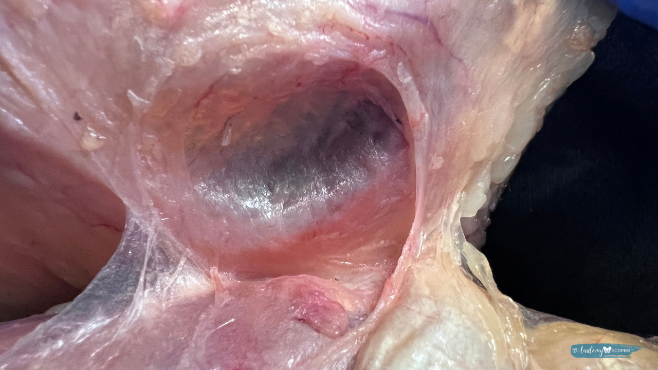
SUBCUTANEOUS BURSA: Subcutaneous bursae (including the olecranon bursa and prepatellar bursa) are found deep to the superficial fascia membrane and exist in the DAT gliding plane. Carla Stecco suggests that they are a specialization of fascia, formed by the fusion of the superficial fascia membrane and deep fascia, creating a virtual space.



⚓️ANCHOR:
Lines of Adhesion & Inframammary Fold
More specializations of the Superficial Fascia! While some superficial fascia-deep fascia interfaces are built to GLIDE, others are built to ANCHOR.
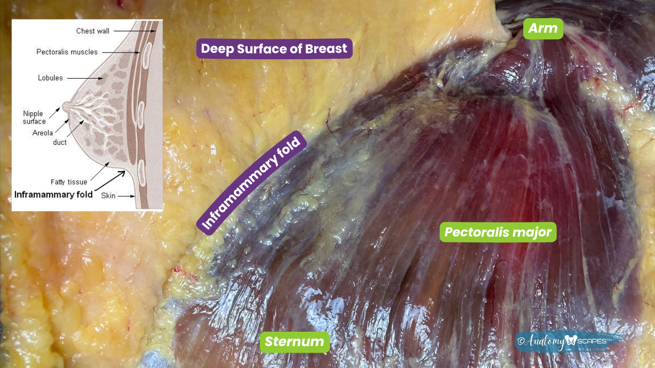
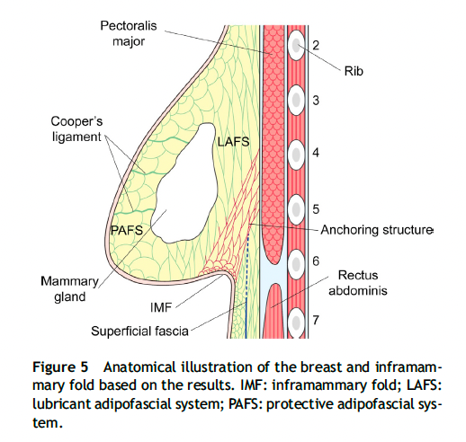
Longitudinal & transverse lines of adhesions exist in specific and consistent locations throughout the human body. In these regions SAT and DAT can be completely absent and instead we find a fusion of the superficial and deep fascia. These lines adhesions are the SAME from person to person.
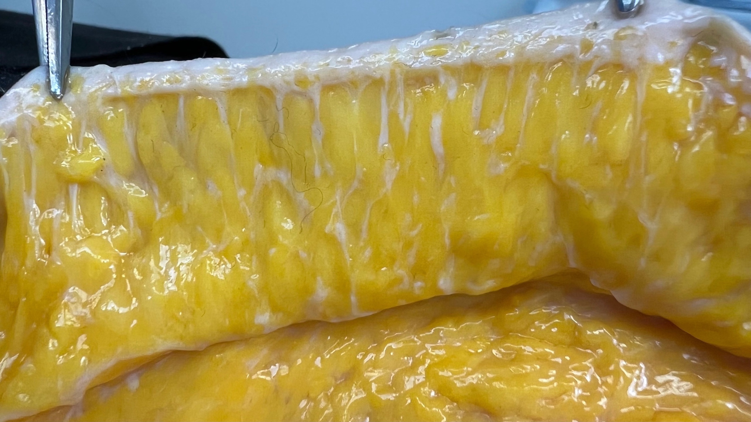
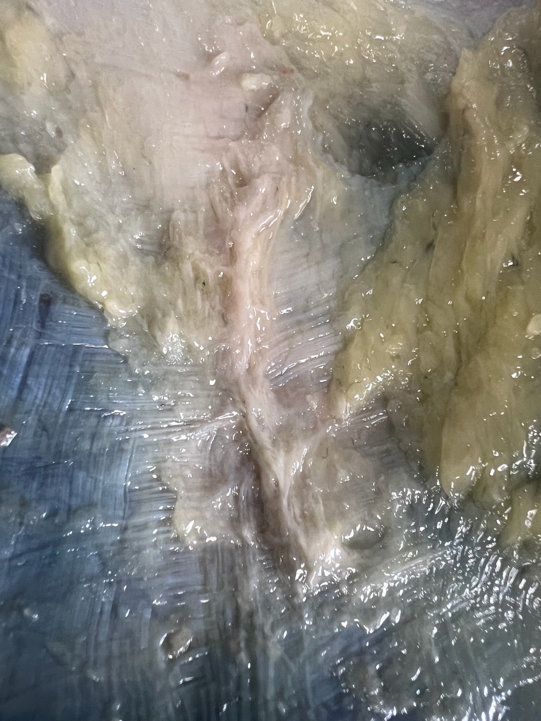
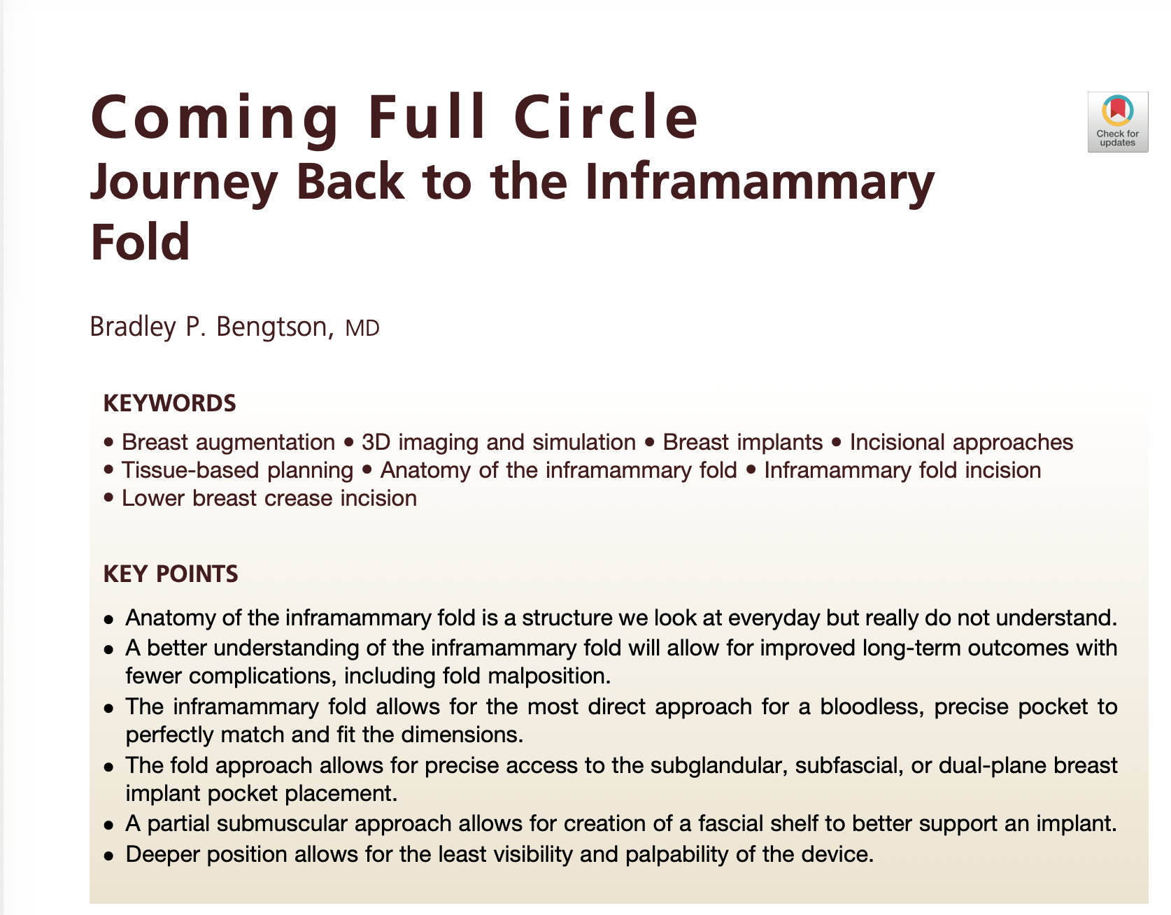
INFRAMAMMARY FOLD: One such transverse line of adhesion is the inframammary fold. (See image of Chelsea's dissection below.) This adhesion between the SF and DF along the sixth rib, forms the The inframammary fold is the curved crease at the lower border of the breast where the skin of the breast meets the skin of the chest wall. Histological workd escribes it as a dense, organized fascial adhesion zone rather than a discrete ligament, though some authors describe a distinct “inframammary ligament.”


DISSECTION & SURGERY VIDEOS
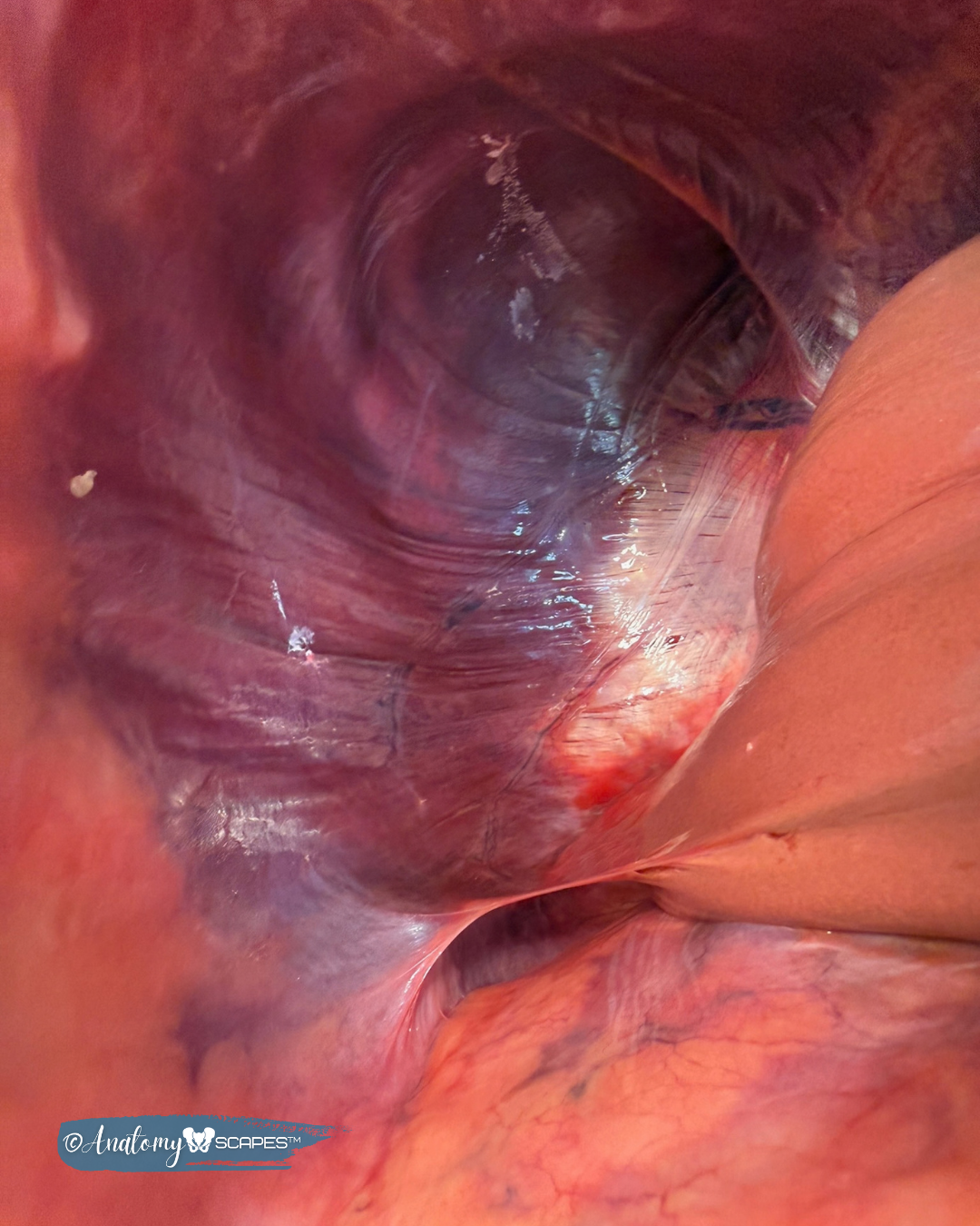
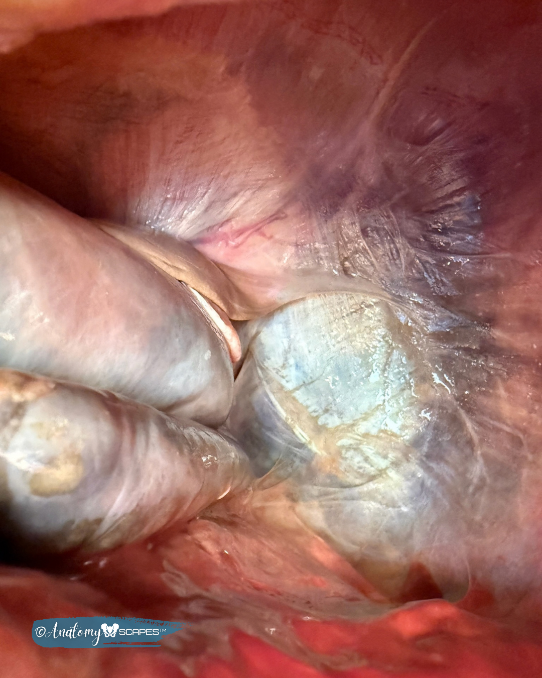
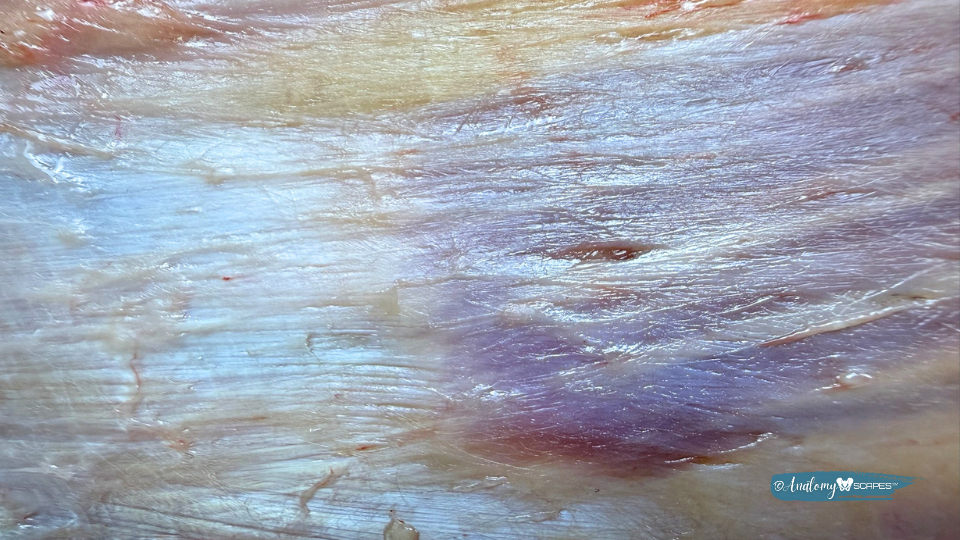
DISSECTION IMAGES
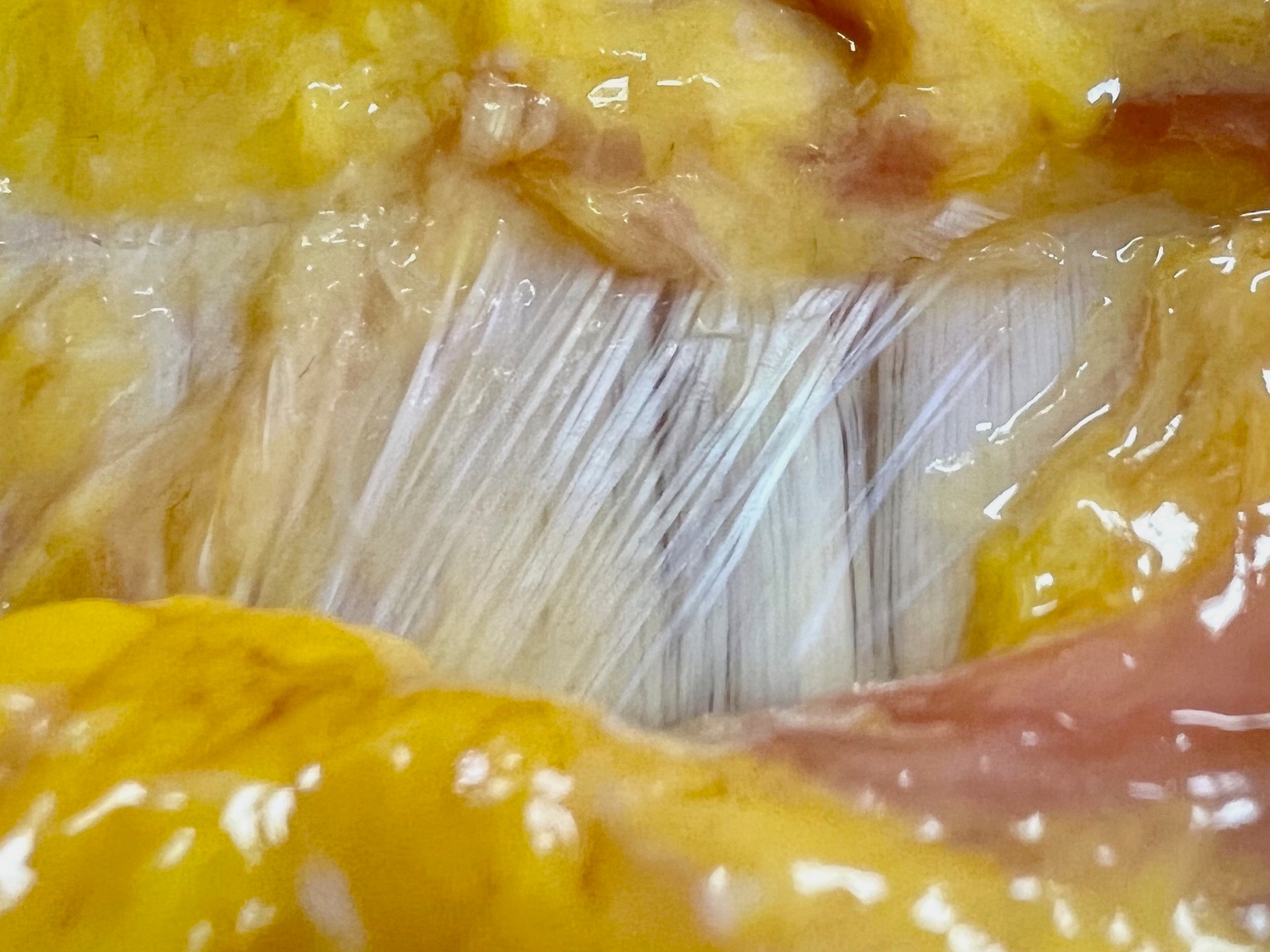
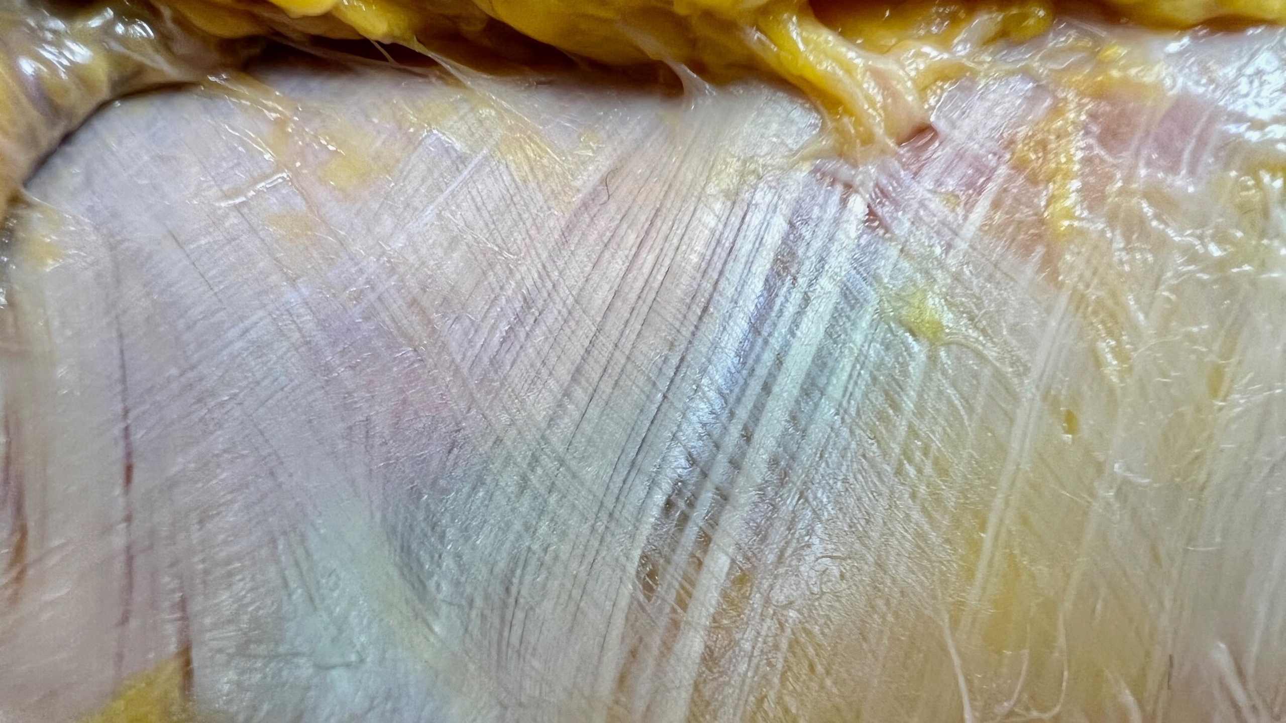
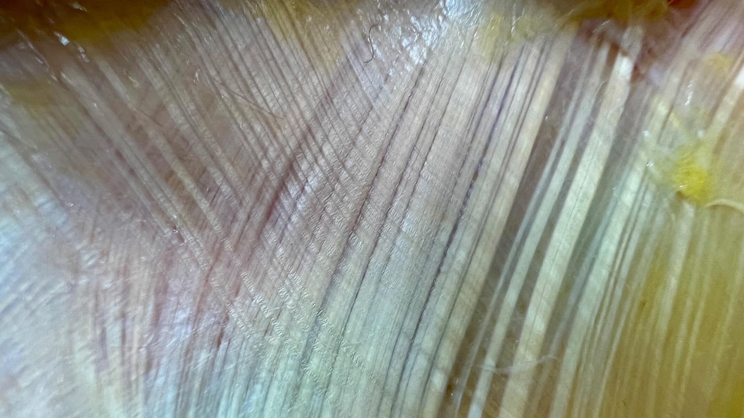
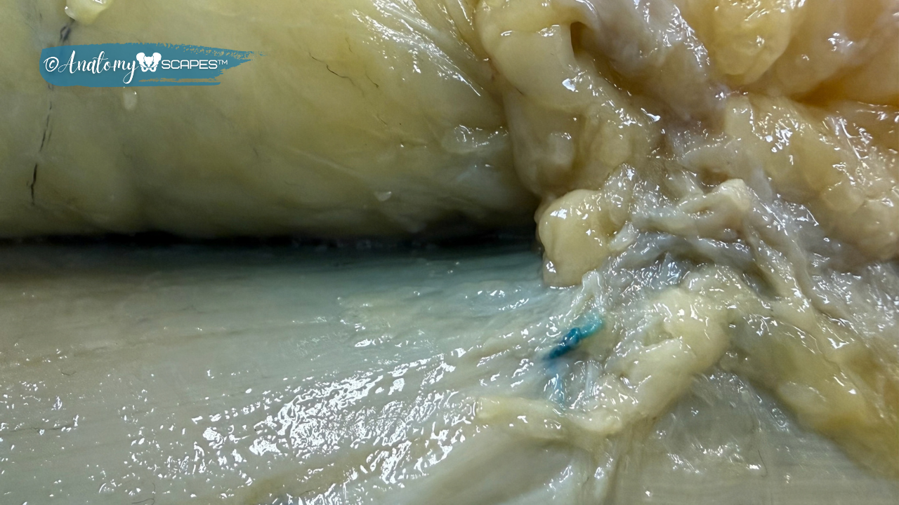
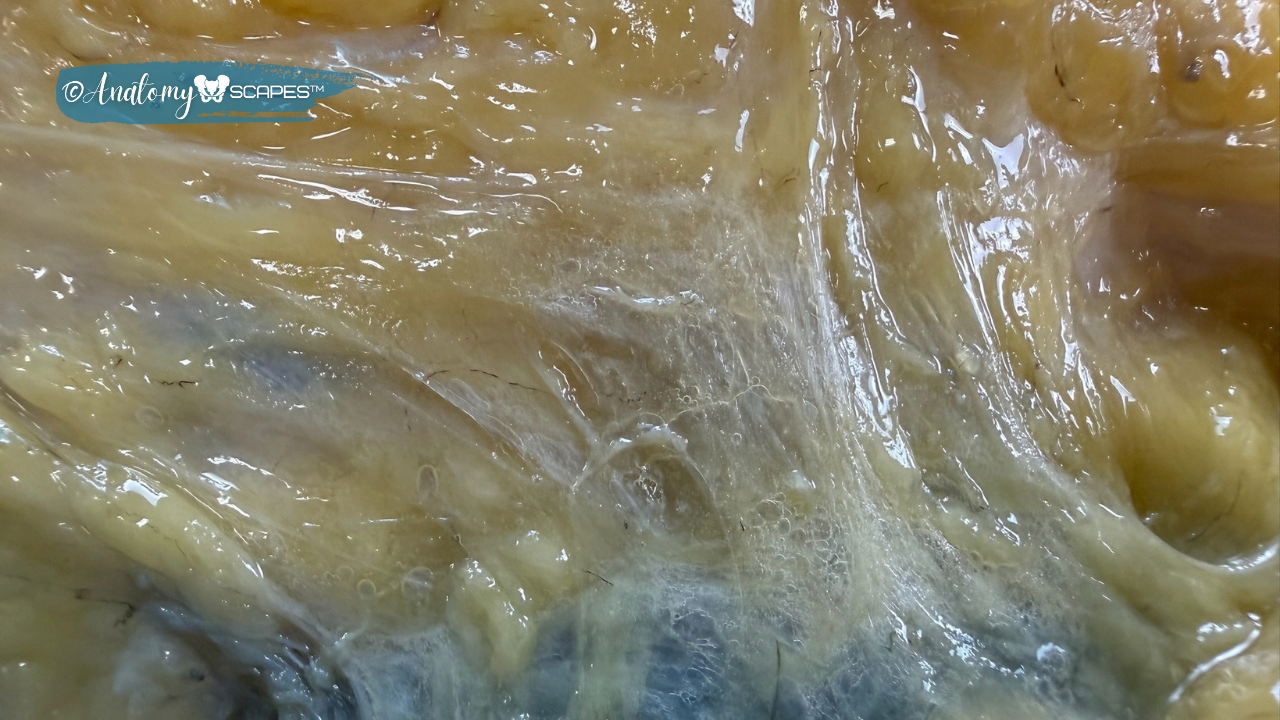
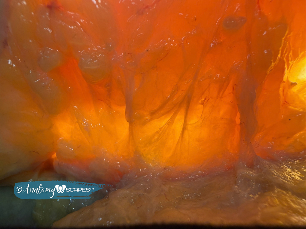
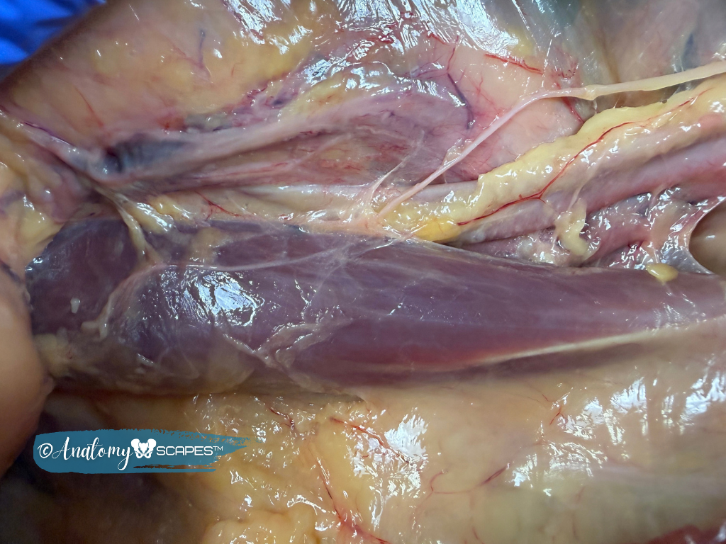
Images from the Dissection Lab
Absolutely NO SCREENSHOTS OR COPYING. AnatomySCAPES' future access to donor programs relies on your responsibility with these materials. The images shown here must remain contained within the educational location you are viewing it with no exceptions.